In-Utero Stem Cell Therapy: A Safe Leap for Fetal Spina Bifida Treatment
Analysis | Technology | March 3, 2026
Key Takeaways
- Safety First: The pioneering Phase 1 trial, known as the CuRe Trial, has demonstrated no significant safety issues, marking a critical green light for further research in fetal regenerative medicine.
- Beyond Standard Surgery: This approach augments traditional fetal surgery for myelomeningocele with a placental stem cell patch, aiming to actively protect and regenerate spinal cord tissue before birth.
- Promising Early Indicators: Initial outcomes, including reversal of hindbrain herniation and no need for postnatal shunts in the first cohort, suggest potential for improved long-term neurological function.
- Regulatory and Ethical Milestone: The FDA's approval to proceed to the next trial phase signals regulatory confidence and opens complex conversations on the ethics of fetal cell-based interventions.
- Broader Implications: Success here could catalyze a new frontier of in-utero therapies for a range of congenital conditions, fundamentally shifting prenatal care paradigms.
A Paradigm Shift in Prenatal Medicine
The landscape of treating severe congenital defects is undergoing a profound transformation. Recent findings from a landmark clinical investigation have confirmed the initial safety of a novel therapeutic strategy that merges advanced fetal surgery with regenerative stem cell science. This intervention, targeting the complex birth anomaly spina bifida, represents not merely a technical achievement but a conceptual leap into the era of active prenatal repair. For decades, the standard of care for the most severe form of spina bifida, myelomeningocele, has involved surgical closure of the spinal defect after birth, or more recently, fetal surgery to cover the exposed tissue. While fetal surgery has improved outcomes, it remains largely protective, not regenerative. The integration of living, placenta-derived stem cells seeks to change that narrative, aiming to shield and potentially repair the developing nervous system within the sanctuary of the womb.
The implications of this successful safety profile extend far beyond a single condition. It validates a foundational premise: that the intrauterine environment can be a therapeutic space for sophisticated cellular and genetic interventions. Historically, the fetus was considered largely inaccessible for direct treatment beyond managing maternal health. This trial, by demonstrating the feasibility and safety of implanting a bio-engineered patch, challenges that historical limitation and paves a regulatory and methodological path for future in-utero gene therapies, tissue engineering, and other regenerative applications for conditions like congenital diaphragmatic hernia, heart defects, and certain metabolic disorders.
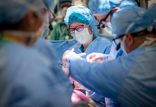
Deconstructing the CuRe Trial: Methodology and Mechanistic Hope
The clinical study, formally titled the "Cellular Therapy for In-Utero Repair of Myelomeningocele (CuRe) Trial," is a meticulously designed first-in-human endeavor. Its Phase 1 primary objective was unequivocal: assess safety. The procedure involves a highly coordinated ballet of maternal-fetal medicine specialists and neurosurgeons. After gaining access to the uterus, surgeons gently position the fetus to expose the spinal lesion. The innovation lies in the application of a patch saturated with mesenchymal stromal cells (MSCs) harvested from donated placental tissue. These cells are not foreign transplants in the traditional sense; they are immunomodulatory and possess trophic properties, meaning they secrete factors that can reduce inflammation, foster a healing microenvironment, and potentially encourage native tissue regeneration.
The choice of placental cells is strategically significant. Unlike embryonic stem cells, they circumvent major ethical debates. Unlike bone marrow-derived cells, they are obtained from medical waste (the placenta) and are thought to be particularly potent due to their perinatal origin. The patch acts as a bioactive scaffold, aiming to do more than just provide a physical barrier. It is intended to interact with the fetal spinal tissue, potentially preventing the progressive damage that occurs from amniotic fluid exposure and mechanical trauma, thereby preserving neurons and neural pathways that would otherwise be lost. The absence of complications like abnormal tissue growth or tumors in the initial six patients is a resounding positive signal for this cellular approach.
Beyond Safety: Analyzing the Unanswered Questions and Future Horizons
While the safety data is cause for optimism, the true measure of this therapy's impact lies ahead. Phase 1 trials are not designed to prove efficacy. The critical questions now transition from "Is it safe?" to "How well does it work?" Future phases must rigorously evaluate long-term functional outcomes in children: mobility, bladder and bowel control, cognitive development, and independence. A key analytical angle missing from early reports is a direct comparison of neurological function at ages 2, 5, and 10 years against matched cohorts who received standard fetal surgery alone. Does the stem cell augmentation lead to measurably stronger leg function, reduced orthopedic surgeries, or improved urological outcomes? These are the metrics that will define success.
Another unexplored dimension is the socio-economic calculus. The current procedure is extraordinarily resource-intensive, requiring a top-tier multidisciplinary team and prolonged hospitalization. What is the cost-benefit analysis if it prevents a lifetime of shunt revisions, wheelchair dependence, and frequent hospitalizations? Health economists must begin modeling this now. Furthermore, the trial raises nuanced ethical considerations. While using placental cells mitigates some concerns, obtaining informed consent for a fetal procedure involving experimental cells is uniquely complex. Parents are making decisions under tremendous emotional duress for two patients—the mother and the fetus—a dynamic that requires exceptional safeguards in the consent process.
The California Catalyst: Funding Innovation at the Frontier
This breakthrough did not occur in a vacuum. It was propelled by a substantial $9 million grant from the California Institute for Regenerative Medicine (CIRM). This investment highlights a crucial model for high-risk, high-reward translational medicine. CIRM's funding bridged the "valley of death" between laboratory research and clinical application, a gap where many promising therapies falter due to lack of capital for the expensive and regulated early-phase human trials. California's continued bet on stem cell research, initially approved by voters in 2004, is now yielding tangible, world-first clinical applications. This success story strengthens the argument for sustained public or public-private investment in regenerative medicine platforms, as the lead time from concept to clinic can span decades.
The CIRM model also suggests that geographic hubs of innovation can develop around such funding. UC Davis Health is now a global epicenter for fetal regenerative surgery, attracting talent and further investment. This concentration of expertise accelerates progress, as seen in the trial's rapid progression to the next phase following the positive safety review by both the FDA and an independent data monitoring board. Their concurrent endorsement is a powerful dual validation of the trial's integrity and promise.
Conclusion: A Cautious Step into a New Era of Prenatal Care
The announcement of safe initial results for in-utero stem cell therapy for spina bifida is a watershed moment in medical technology. It cautiously opens a door that was previously only a theoretical possibility. The path forward remains long and must be tread with scientific rigor and ethical vigilance. Subsequent trial phases will demand meticulous long-term follow-up to ensure no delayed adverse effects emerge and to quantify the genuine functional benefits for children and their families.
Nevertheless, the significance is undeniable. We are witnessing the embryonic stages of a field—fetal regenerative medicine—that could redefine how society addresses congenital disease. Instead of solely managing lifelong disabilities, medicine is inching toward the possibility of mitigating them before birth. As Principal Investigator Diana Farmer suggested, the future is indeed exciting for cell and gene therapy before birth. This trial is the bold, first step in translating that excitement into a new standard of hope and healing for generations to come.
Glossary of Key Terms
Myelomeningocele: The most severe form of spina bifida, where the spinal canal and membranes protrude through an opening in the vertebrae.
Mesenchymal Stromal Cells (MSCs): Multipotent stem cells found in various tissues, including placenta, known for their anti-inflammatory and tissue-supporting properties.
Hindbrain Herniation (Chiari II Malformation): A complication of spina bifida where the brainstem extends into the spinal canal; its reversal is a positive sign of reduced spinal fluid pressure.
Hydrocephalus: A buildup of cerebrospinal fluid in the brain, often requiring a shunt for drainage, common in severe spina bifida.
Phase 1 Clinical Trial: The first stage of testing in humans, primarily focused on evaluating safety and dosage, typically involving a small number of participants.